Microscopy

Microscopy

The scientific service group microscopy provides resources, technology, and expertise to study the morphology and function of cells, tissues, and organisms. Supported imaging technologies range from the visualization of molecules, cellular light and electron microscopy to automated scanning and analysis of hundreds of microscope slides. We teach all the technology we offer as a service, continuously evaluate new technology, establish novel application workflows, carry out quality control, maintain facility instruments, consult users on experimental design, offer advice on data management, and provide limited support for image analysis.
Strategic Plan

Strategic Plan

Global cellular organization shapes and constrains organ function; alterations in cellular networks are of high diagnostic and prognostic value. Therefore, we plan to establish a quantitative framework to capture, analyze and compare the organization of organs using light and electron microscopy. To this aim we have initiated the following Service Pilots: Quantitative Semi-Automated EM, which aims at automating the time-consuming task of manual data acquisition in electron microscopy, and Large High-resolution Scans, 3D Whole Embryo Reconstruction, and Towards Full Automation of Image Capture and Processing, which together pave the way for the establishment of a digital pathology platform. Please contact us if you want to learn more about these projects.
Sample preparation and handling

Sample preparation and handling

Sample preparation prior to imaging, and sample handling during image acquisition are important to obtaining true and reproducible results. Post-mortem changes in tissues during EM sample preparation, issue related to antibody staining, artifact produced by the use of aggregating genetically encoded fluorophores and cellular stress, induced during in vivo imaging, are frequent causes of misleading or non-reproducible results. Thus, some workflows, such as the embedding of samples for electron microscopy are at current primarily offered as full service. For critical parts of the sample preparation pipeline performed by the scientist, such as perfusion prior to EM embedding, and for keeping tissues alive during in vivo imaging, reliable protocols and detailed instructions on how to quality control the experiment are provided. For all quantitative biology projects, the entire workflow will be planned jointly. Instruments: Leica EM TP (tissue processor), Leica EM TRIM2 (milling system), Reichert Jung Ultracut E (ultramicrotome), environmental chambers at all confocal imaging systems.
Electron Microscopy

Electron Microscopy

We provide full service and in-depth practical training in transmission electron microscopy. We recently developed a workflow that allows for capturing large regions of interest semi-automatically. Service Pipelines: Negative Stain & Qualitative cellular EM Service Pilot: Quantitative Cellular EM, Semi-Automated & Adaptive Feedback EM* (*only in collaboration) Instruments: JEOL JEM 1400 Plus (120 keV electron microscope)
Light Microscopy

Light Microscopy

In general, light microscopes are operated by the user after initial training. We offer advice and support for experimental design, sample preparation, and staining, as well as on how to maintain tissue and organism alive during long-term imaging. Projects at the slidescanner are either offered as full service or in supervised use. Instruments: Leica SP8 MP (multi-photon microscope), Zeiss 880 Airyscan (confocal microscope), Nikon Ti-E with Märzhäuser SlideExpress 2 (slidescanner) Service Pipelines: Large High-Resolution Scans

High-resolution scan of a hematoxylin and eosin (HE) stained renal paraffin section. The resulting image (A) is composed of more than 500 individual images acquired with a 40x 0.95 NA Air objective. Selected regions (white boxes in A,B) are shown enlarged in (B,C). (C) Connectivity of individual cells can be clearly distinguished. Scale bars:  (A): 800 µm, (B) 200µm,(C): 20µm
Quality Management

Quality Management

We regularly assure the quality of our instruments and services and improve our application workflows continuously. Quality management is for us a mindset that guides all our actions. Please contact us personally, if you want to learn more about our procedures and tools, or if your project requires certification according to a certain norm.

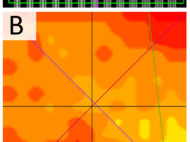
Example of results of quality checks performed to control the performance of our instruments. (A) A pattern of gradually spaced lines is scanned to asses the lateral resolving power. (B) A normalized homogeneity map used to quantify illumination inhomogeneity.

External links

 Microscopy primer all about microscopy Confocal Listserver Discussion forum for microscopy subjects image.sc Discussion forum for image analysis subjects Societies, Initiatives & Networks: ELMI The European Light Microscopy Initiative is a communication network between European scientists working in the field of light microscopy and the manufacturers of their equipment. Euro-BioImaging Research Infrastructure for Imaging Technologies in Biological and Biomedical Sciences DGE German Society for Electron Microscopy German BioImaging Biological Imaging Facilities in Germany neubias Network of European BioImage Analysts
Go to Editor View